Difference between revisions of "Zeiss Axio Imager M2m Microscopes"
Jump to navigation
Jump to search
(update tool owner, update to NEMO) |
|||
| Line 8: | Line 8: | ||
| Instrument_Type = Metrology | | Instrument_Type = Metrology | ||
| Staff_Manager = [[Kyle Keenan | Kyle Keenan]] | | Staff_Manager = [[Kyle Keenan | Kyle Keenan]] | ||
| − | | Lab_Location = | + | | Lab_Location = Cleanroom Corridor |
| Tool_Manufacturer = Zeiss | | Tool_Manufacturer = Zeiss | ||
| Tool_Model = Axio Imager M2m | | Tool_Model = Axio Imager M2m | ||
Latest revision as of 11:45, 28 June 2024
 |
|
| Tool Name | Zeiss Axio Imager M2m Microscopes |
|---|---|
| Instrument Type | Metrology |
| Staff Manager | Kyle Keenan |
| Lab Location | Cleanroom Corridor |
| Tool Manufacturer | Zeiss |
| Tool Model | Axio Imager M2m |
| NEMO Designation | MET-12, MET-13, MET-14, MET-15 |
| Lab Phone | XXXXX |
| SOP Link | SOP |
Description
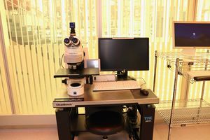
In Normal operation, it is possible to observe ~2 µm diameter objects in the reflected and transmitted mode. Furthermore, the following contrasting techniques are available: bright-field, dark-field, Circular Differential Interference Contrast (C-DIC) using circularly polarized light, polarization contrast, and polarization with additional retarder. The image snapped can be analyzed using the annotation tool on the software.
In Extended Focus operation, the images of 3D structure can be acquired at different focus positions, and automatically combined as a sharp 2D image.
In Panorama operation, the images acquired can be stitched, so that the optical microscope image of the wide object can be obtained.
Applications
- Bright-field Imaging
- Dark-field Imaging
- Circular Differential Interference Contrast (C-DIC) Imaging
- Polarization contrast Imaging